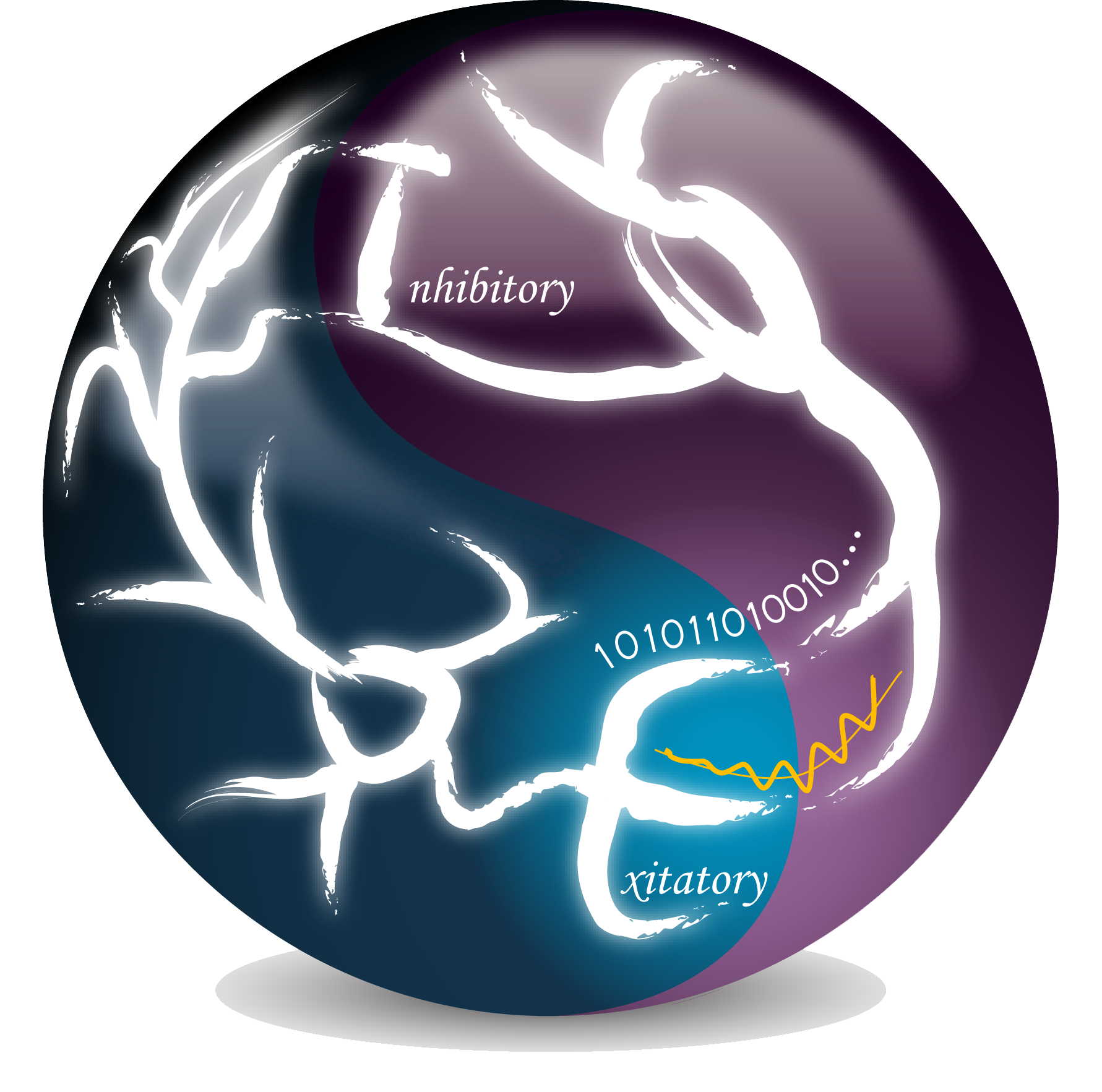
We developed a method of tight-seal axon recording (Shu et al., 2006 Nature; Shu et al., 2007 J. Neurophysiol.). After obtaining whole-cell recording from the soma with a patch pipette filled with fluorescent dye, we trace the axon down to the cut end where the axon membrane reseal and form a bleb. This bleb has a diameter of 3-5 mm, a perfect site for axon recording. Another patch pipette (7-15 MOhm) usually containing no dye is used for bleb recording. Therefore, simultaneous somatic and axonal whole-cell recording, regular outside-out or inside-out patch recording, or isolated bleb recording can be achieved (Hu et al., 2009 Nature Neurosci.; Hu and Shu, 2012 Neurosci. Bul.).
To obtain isolated bleb recording, we sweep a sharp electrode at the border between layer 6 and white matter, then choose to record axon blebs at the white matter side. Loading the bleb with fluorescent dye allows us to determine whether the bleb is disconnected from the somatodendritic compartment (Left figure). Using this method, we can obtain pure axonal currents. Our results suggest that the axon express dopaminergic receptors and activation of these receptors may modulate axonal K
+ currents and consequently regulate the waveform of axonal action potentials (Yang et al., 2013 J Physiol.).
